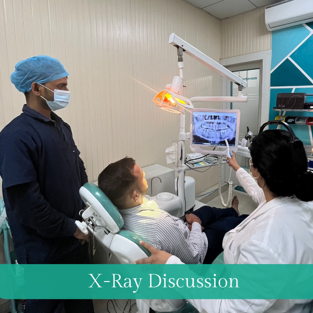
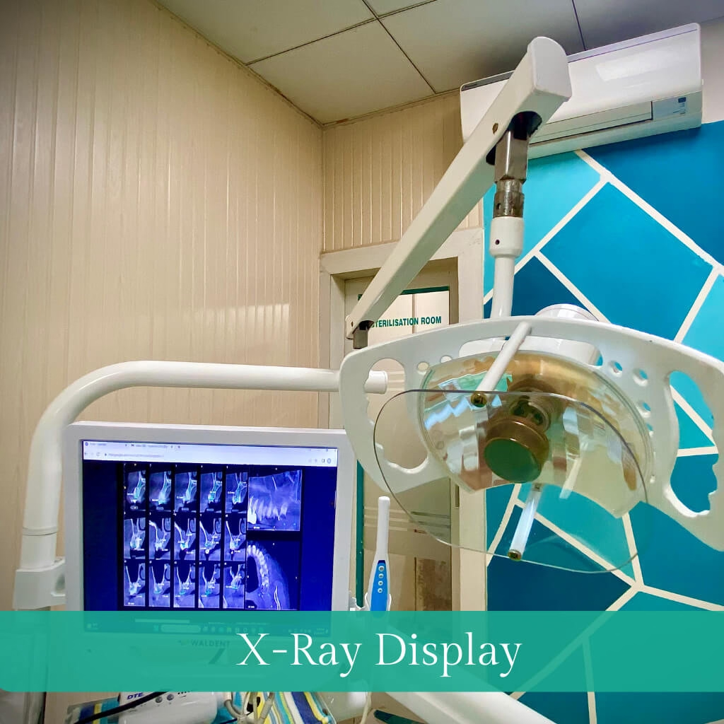
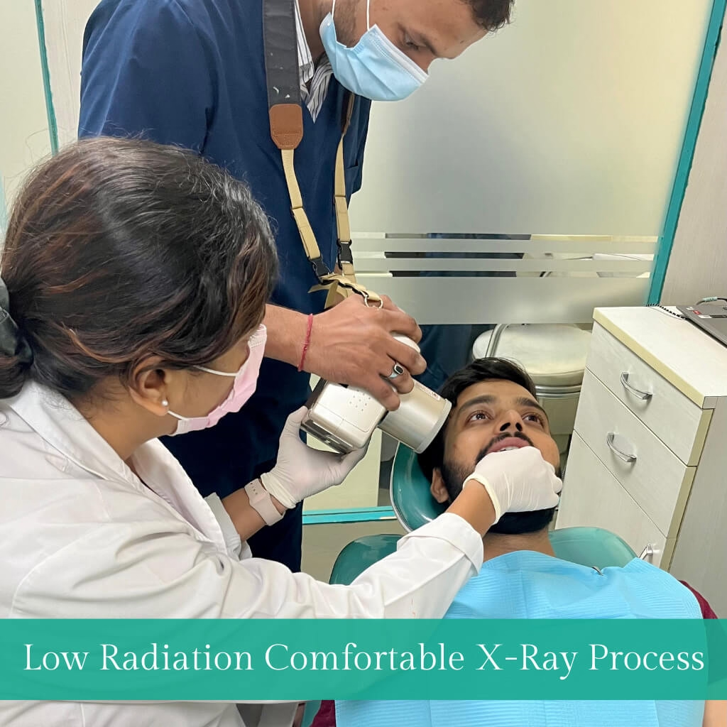
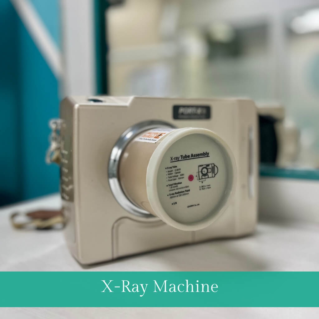
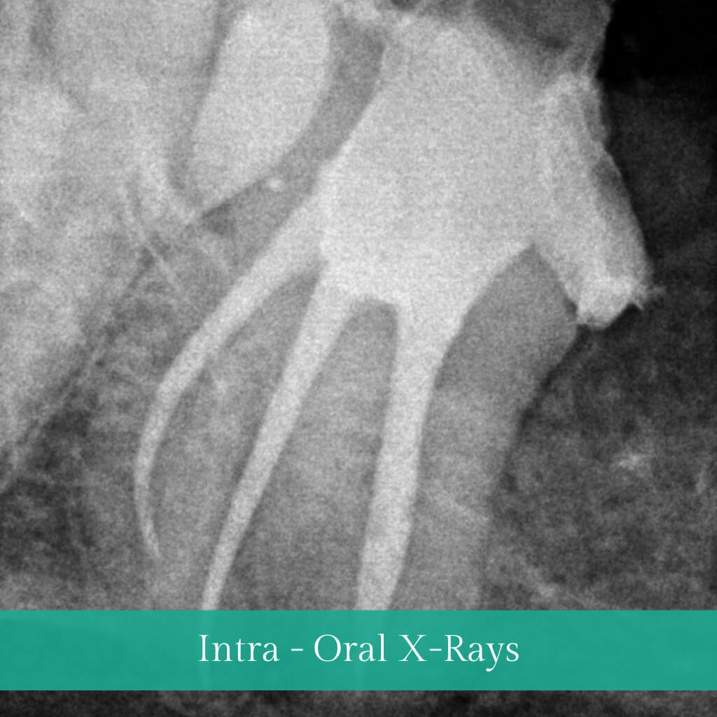
Dental X-rays, also known as dental radiographs, are diagnostic imaging techniques used in dentistry to visualise and assess various structures within the oral cavity, teeth, and surrounding tissues. These X-rays provide valuable insights that are not visible during a regular dental examination. Dental X-rays are essential tools for detecting dental issues such as cavities, gum diseases, impacted teeth, bone abnormalities, and structural problems.
They play a crucial role in diagnosis, treatment planning, and ongoing monitoring of oral health, helping dental professionals provide effective and personalised care. Established protocols guide the use of dental X-rays to minimise radiation exposure while maximising diagnostic benefits.
What are the types of Dental X-Rays?
Bitewing X-rays
Used to detect cavities between teeth and assess dental crowns' fit.
Periapical X-rays
Provide detailed views of individual teeth from Crown to root, aiding in diagnosing dental issues.
Panoramic X-rays
Capture a broad view of the mouth, including jaws and surrounding structures.
Orthodontic X-rays
Required for orthodontic treatment planning, showing tooth positioning and growth. Cephalometric X-rays are used in orthodontics to assess facial structures, especially concerning the teeth and jaws.
Cone Beam Computed Tomography (CBCT)
Offers advanced 3D imaging for comprehensive assessments, commonly employed in implant planning and intricate dental cases.
What can Dental X-Rays detect?
- Cavities (dental caries)
- Impacted or unerupted teeth
- Tooth and root infections (abscesses)
- Gum diseases (periodontal diseases)
- Bone loss in the jaw
- Jaw fractures or abnormalities
- Dental cysts or tumours
- Root canal issues
- Dental decay beneath existing fillings
- Positioning of teeth and jaw alignment for orthodontic treatment planning.
Is radiation from Dental X-rays harmful?
No form of radiation is desirable for an individual; however, taking certain measures can minimise or even eliminate the risk of radiation exposure for patients. We at Hope Dental and Esthetic Clinic use only AERB-approved machines with minimum radiation scatter making them safe for our patients and staff. The utilisation of Lead aprons and thyroid collars are other adjuncts we employ to minimise the risk of radiation-associated concerns.


